
Foot & Ankle Pain Treatment in Bend, Oregon
At High Desert Sports & Spine, our fellowship-trained, board-certified physicians evaluate and treat foot & ankle pain in Bend, Oregon, with a focus on non-operative and interventional options.
Foot and ankle pain has a particular way of wearing people down. You feel it in the first steps out of bed, and again at the end of a shift, and again on the trail you used to run without thinking about it. Most people put up with it far longer than they would with pain anywhere else.
Much of that pain comes from degenerated tendons and fascia rather than inflammation, which is why rest and anti-inflammatories often help a little and then stop helping. The tissue needs to rebuild, and that takes a different approach than waiting it out.
At High Desert Sports & Spine, our fellowship-trained, board-certified physicians evaluate and treat foot and ankle pain in Bend, Oregon, using in-office ultrasound and interventional treatment including shockwave therapy, image-guided injection, and orthobiologics. We look at the tissue directly, tell you what is actually going on, and build a plan around it.
Same-week appointments are usually available for new patients.


When Foot or Ankle Pain Needs a Specialist
Consider a specialty evaluation if you are dealing with:
- Heel pain that is worst with the first steps in the morning and has lasted more than 6 weeks
- Pain in the back of the heel or above it that has not responded to stretching and rest
- An ankle that keeps rolling, or that feels unreliable on uneven ground
- An ankle sprain that has not fully settled after several weeks
- Pain along the inside of the ankle and arch, particularly if your arch looks like it is flattening
- Burning, numbness, or tingling in the foot
- Pain that gets worse the further you run or walk
- Persistent pain after foot or ankle surgery
If you have diabetes and notice a sore, blister, non-healing wound, or any area of redness, warmth, or drainage on your foot, do not wait for a routine appointment. Diabetic foot wounds can progress quickly and need prompt evaluation. Contact your primary care provider, a podiatrist, or seek urgent care the same day.
For an acute foot or ankle injury, our Orthopedic Urgent Care offers same-day or next-day evaluation without the ER wait.
Foot & Ankle Conditions We Evaluate and Treat in Bend, OR
Plantar Fasciitis and Heel Pain
The signature of plantar fasciitis is sharp heel pain with the first steps in the morning, easing as you move and then returning after you sit for a while or at the end of a long day on your feet.
The name suggests inflammation, and for a long time that is how it was treated. Tissue studies changed the picture. Chronic plantar fascia pain shows degeneration and disorganized tissue rather than active inflammation, which is why anti-inflammatories and rest so often produce partial, temporary relief. The fascia has broken down and has not rebuilt itself.
That changes what works. Shockwave therapy has some of the strongest evidence of any treatment in this area, with systematic reviews finding meaningful effects on both pain and function that hold up over time. It requires no downtime and no needles. We use both focused and radial shockwave, which lets us match the modality to what the ultrasound shows. We use radial shockwave for the calf and surrounding areas, while focused shockwave targets the plantar fascia directly.
Ultrasound also lets us measure fascia thickness and confirm the diagnosis rather than assume it, which matters because heel pain has several other causes. A heel fat pad problem, a calcaneal stress fracture, and entrapment of a small nerve near the heel all produce pain in roughly the same place and do not respond to plantar fascia treatment.
A word on cortisone. Steroid injection into the plantar fascia can provide real short-term relief, and there are situations where that is worth it. It also carries documented risks, including rupture of the fascia and atrophy of the heel fat pad, the natural cushion under your heel that does not grow back. We use it selectively and will explain the tradeoff rather than leading with it.
Heel spurs, while we are here, are usually incidental. Plenty of people have them with no pain at all, and the spur is rarely what needs treating.
Achilles Tendinopathy
Achilles problems come in two varieties, and they behave differently, so it’s worth making the distinction.
Midportion Achilles tendinopathy affects the tendon a few centimeters above the heel bone. Insertional Achilles tendinopathy affects the point where the tendon attaches to the heel, and it is generally more stubborn and less tolerant of the stretching that helps the midportion kind.
Both cause pain and stiffness that are worst first thing in the morning and at the start of activity, sometimes easing as you warm up and then returning afterward. Runners, hikers, and anyone who has recently increased their volume or changed their footwear are the usual candidates.
Progressive loading is the foundation of treatment and has the best evidence behind it. We coordinate closely with physical therapists across Central Oregon who do this well. Ultrasound lets us confirm which part of the tendon is involved and check for partial tearing, which changes how aggressively we can load the tendon. For cases that stall despite good rehabilitation, options may include shockwave therapy, PRP, or percutaneous tenotomy.
There is good evidence for shockwave for mid-substance Achilles tendon, and we use it regularly as a first-line treatment. We also avoid corticosteroid injection into or around the Achilles because the tendon carries your full body weight with every step, and steroid injection is associated with rupture risk. That is a case where short-term relief isn’t worth what it may cost you.
Posterior Tibial Tendinopathy
The posterior tibial tendon runs behind the inner ankle bone and holds up the arch of your foot. When it becomes irritated or begins to fail, pain develops along the inside of the ankle and arch, often with swelling, and it gets worse the longer you are on your feet.
This one matters more than most people realize, because posterior tibial tendon dysfunction is progressive. As the tendon stretches and weakens, the arch gradually collapses, and the foot changes shape, and the later stages are considerably harder to manage than the early ones. A useful early sign is difficulty rising onto the toes of one foot, or noticing that one arch looks flatter than the other.
Ultrasound lets us assess the tendon directly and identify thickening or tearing. Treatment centers on supporting the arch, unloading the tendon, and a targeted strengthening program, with image-guided injection or orthobiologic treatment considered in appropriate cases. Catching this early genuinely changes the outcome, which is the reason it gets its own section.
Ankle Sprains, Strains, and Ligament Tears
Ankle sprains are the most common musculoskeletal injury, and they are also the most commonly dismissed. The phrase “just a sprain” does a lot of damage, because a meaningful share of people who sprain an ankle go on to have ongoing symptoms or recurrent instability, often because the first injury was never properly rehabilitated.
Most sprains involve the ligaments on the outside of the ankle from a rolling-inward injury. High ankle sprains, which affect the ligaments connecting the two lower leg bones, are less common, take considerably longer to heal, and are frequently missed early.
Early evaluation is worth more here than people expect. We use examination, ultrasound, and on-site X-ray to grade the injury and rule out fracture, and ultrasound lets us assess the ligaments dynamically rather than just at rest. Getting the grade right sets the rehabilitation timeline, and a proper rehabilitation program is the main thing standing between a sprained ankle and a chronically unstable one.
Chronic Ankle Instability
If your ankle keeps rolling, feels unreliable on trails or uneven ground, or you find yourself watching the ground instead of the view, you may have chronic ankle instability.
It develops in two ways, and usually both at once. The ligaments may have healed loose or lengthened after previous sprains, and injury can disrupt the nerve feedback system that tells your brain where your foot is in space. That second part is often the bigger problem, and it also responds best to training.
We assess ligament integrity with examination and dynamic ultrasound, evaluate the peroneal tendons along the outside of the ankle since they are commonly involved, and look for what often gets missed in repeatedly sprained ankles, including small fractures and cartilage injuries inside the joint. Most cases respond to a structured balance and strengthening program. Ankles with significant ligament laxity that continue to give way despite good rehabilitation may need surgical stabilization, and we coordinate that evaluation.
Foot and Ankle Arthritis
Foot and ankle arthritis causes stiffness, aching, and swelling that is typically worst in the morning and after activity. The ankle joint, the midfoot joints, and the big toe joint are common sites. Ankle arthritis frequently follows an old fracture or a history of repeated sprains, sometimes decades later.
These are small joints and difficult targets, which is why image guidance matters. Ultrasound-guided or X-ray-guided injection can provide meaningful relief and also helps confirm which joint is actually responsible when several look abnormal on X-ray. For patients with early to moderate arthritis, orthobiologic treatment may be worth discussing. Footwear modification and load management often do more than patients expect.
Nerve Pain, Neuropathy, and Nerve Entrapment
Burning, tingling, numbness, or electric pain in the foot points to a nerve rather than a joint or tendon. What is causing it matters, because the treatments are entirely different.
Peripheral neuropathy, including diabetic neuropathy, typically affects both feet in a symmetric pattern, often starting in the toes and moving upward, and is frequently worse at night. It is a medical condition, and your primary care provider or a neurologist manages it. We do not provide blood sugar control, medication management, or diabetic foot care, including wound and ulcer treatment, and we are not going to pretend otherwise. A lot of marketing in this space promises to reverse neuropathy. We encourage you to be skeptical.
What we can contribute is a different question: whether your foot pain comes from a focal nerve entrapment rather than diffuse neuropathy. These are mechanical problems, and they are treatable.
Tarsal tunnel syndrome involves compression of the tibial nerve behind the inner ankle bone, producing burning or tingling in the sole of the foot. Entrapment of the first branch of the lateral plantar nerve is a commonly missed cause of heel pain that can be mistaken for plantar fasciitis and may not respond to plantar fascia treatment. Morton’s neuroma involves an irritated nerve between the toes and causes burning forefoot pain, often with a pebble-in-the-shoe sensation.
Ultrasound can evaluate all three, and treatment may include an image-guided injection or hydrodissection, which uses precisely placed fluid to free a nerve from the tissue restricting it. Patients with diabetes can have both diffuse neuropathy and a focal entrapment at the same time, and separating the two is a genuinely useful thing to sort out.
Morton’s Neuroma
Morton’s neuroma is an irritated and thickened nerve between the toes, most often between the third and fourth. It causes burning or sharp pain in the ball of the foot, sometimes with numbness in the toes, and patients frequently describe the sensation of a pebble in the shoe or a sock bunched up underneath.
Narrow or tight footwear is a common contributor, and symptoms often ease when you take the shoe off and rub the foot. It is more common in runners and in people who spend long days on hard surfaces.
Ultrasound is well suited to this. We can see the nerve directly, measure it, and confirm the diagnosis rather than relying on symptoms alone, which matters because stress fractures and forefoot joint problems can cause similar pain. Treatment starts with footwear changes and offloading. For persistent symptoms, ultrasound-guided injection or hydrodissection can reduce irritation and free the nerve from surrounding tissue. Cases that do not respond may be candidates for surgical excision, and we coordinate that referral.
Shin Splints and Lower Leg Pain
Medial tibial stress syndrome, commonly called shin splints, causes aching pain along the inner edge of the shin that shows up during running and eases with rest. It usually follows a jump in training volume, a change in surface, or a return to running after time off.
The important distinction is between shin splints and a tibial stress fracture, since the management differs substantially. Shin splints typically produce pain spread along a stretch of the bone that improves as you warm up. A stress fracture tends to hurt at one specific point, gets progressively worse rather than improving with warming up, and eventually hurts with walking or at night. Early X-rays are often normal in stress fractures, so they can be missed and left untreated for weeks.
We evaluate with examination, on-site X-ray, and MRI referral when a stress fracture is suspected, and we look at why the bone or tissue failed in the first place, including training load and mechanics. That last part helps prevent the next one.
Stress Fractures of the Foot and Ankle
Stress fractures in the foot are common in runners, hikers, and anyone who increases their mileage too quickly. The metatarsals are the usual site, along with the navicular in the midfoot and the calcaneus in the heel.
The pattern is localized pain that builds with activity and eventually persists at rest. Two locations warrant extra caution: navicular stress fractures and stress fractures at the base of the fifth metatarsal both have a reputation for poor healing and sometimes need surgical fixation or prolonged protected weight bearing.
We evaluate with on-site X-ray and coordinate MRI promptly when the clinical picture suggests a stress fracture despite normal films. Most heal well with protected weight bearing and a structured return to activity, and we look at contributing factors including training load, footwear, and bone health.
Chronic Pain After Foot or Ankle Surgery
Persistent pain after foot or ankle surgery, including fracture fixation, ligament reconstruction, and bunion or fusion procedures, is more common than most patients are told to expect. Scar tissue, nerve entrapment in the surgical field, altered mechanics, and problems that were never the original target all contribute.
A specialty evaluation including your operative reports and prior imaging helps clarify what is generating the pain now. Diagnostic blocks are particularly useful here because they can isolate a specific structure when surgery has changed the anatomy, and ultrasound-guided hydrodissection is an option when a nerve is tethered in scar tissue.









How We Diagnose Foot & Ankle Pain
The foot and ankle pack twenty-six bones, thirty-three joints, and a great many tendons and nerves into a small space. Several structures can cause pain in nearly the same spot, so evaluation is the most important part.
A typical evaluation includes:
- History and symptom review. When it started, whether there was a specific injury, whether it is worse with the first steps of the day or at the end of it, what surfaces and shoes make it worse, and what you are trying to get back to. Morning-first-step pain points somewhere different than pain that builds through a run.
- Physical examination. We assess range of motion, strength, ligament stability, nerve function, and gait. We look at the whole chain, since hip and knee mechanics change how the foot is loaded and vice versa.
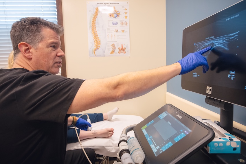
- In-office musculoskeletal ultrasound. The foot and ankle are close to the surface, which makes them among the best regions in the body for ultrasound. We can see the plantar fascia, Achilles and posterior tibial tendons, ankle ligaments, and nerves in real time, and assess ligaments dynamically as the ankle moves. Much of the diagnosis happens in the room.
- On-site X-ray. Available at the same visit to evaluate bone, joint space, alignment, and suspected fracture.
- MRI referral. Arrange when a stress fracture, cartilage injury, or significant tendon tear needs characterization.
Foot & Ankle Treatment Options at HDSS
Shockwave Therapy (ESWT)
Acoustic energy is delivered to degenerated tissue to stimulate healing. Plantar fasciitis is one of the strongest applications for shockwave anywhere in musculoskeletal medicine, supported by high-quality evidence for both pain and function. We offer both focused and radial shockwave, performed in the office with no downtime and no needles. Most insurance providers do not cover shockwave treatments, and our team will go over the cost before anything is scheduled.
Ultrasound-Guided Injections
We perform every injection under real-time ultrasound or fluoroscopy. In the foot and ankle, the targets are small and sit close to nerves, tendons, and the fat pad under the heel, so accuracy affects both whether the treatment works and whether it causes harm.
Nerve Hydrodissection
Precisely placed fluid separates an irritated or tethered nerve from the tissue restricting it. Relevant for tarsal tunnel syndrome, heel nerve entrapment, Morton’s neuroma, and nerve adhesions after foot or ankle surgery.
PRP Therapy
Platelet-rich plasma delivered under ultrasound guidance into degenerated fascia or tendon. Most relevant for chronic plantar fasciitis and Achilles tendinopathy that have not responded to loading and shockwave. Results develop over weeks to months.
Percutaneous Tenotomy
For tissue with established degeneration, a small ultrasound-guided needle breaks up disorganized tissue and prompts a healing response.
MLS Laser Therapy
Non-invasive light therapy to reduce inflammation and support recovery, used as an adjunct alongside other treatment.
Bracing, Footwear, and Load Management
A change in footwear, a heel lift, an ankle brace, or an adjustment to training volume is frequently the highest-value part of the plan. We address footwear and how you are loading the foot as part of the plan, and we refer to podiatry when custom orthotics are the right call.
Rehabilitation Coordination
For tendon and fascia problems, progressive loading has the strongest evidence, and for ankle instability, balance and strength training is the primary intervention. We build these into the plan and coordinate with physical therapists across Central Oregon.
Coordination of Care
Unstable fractures, ligament injuries that need surgical stabilization, advanced arthritis, and conditions outside our lane, including bunions, hammertoes, nail and skin problems, and diabetic wound care are referred to the right specialist. We have good working relationships with podiatry and orthopedic surgery in Central Oregon and will point you in the right direction rather than treating something we are not the best fit for.


Who May Benefit from Specialty Foot & Ankle Care
An evaluation at High Desert Sports & Spine may be worth considering if you:
- Have had heel pain for months despite stretching, inserts, and rest
- Were offered a cortisone shot for heel pain and want to understand the alternatives first
- Have Achilles pain that keeps coming back every time you return to running
- Have an ankle that rolls repeatedly or feels unreliable on trails
- Have pain along the inside of your ankle and have noticed your arch changing
- Have burning or tingling in your foot and want to know whether something treatable is contributing
- Have pain that has persisted after foot or ankle surgery
- Are a runner or hiker with pain that is getting worse rather than settling
What to Expect at Your Foot & Ankle Evaluation
- Specialty evaluation. History, prior treatment, any imaging you have, your footwear, your training, and what you are trying to get back to.
- Hands-on exam and in-office imaging. Physical examination plus ultrasound or X-ray as indicated, during the same visit.
- Clear diagnosis and options. You will understand which structure is involved, what the evidence supports, and what a realistic timeline looks like. Tendon and fascia problems take months rather than weeks, and we will say so up front.
- A plan you agree with. Including the loading and footwear side, not just the procedural side. No pressure and no upsell.
- Follow-up. We track your response and adjust, and coordinate rehabilitation throughout.


Why Choose High Desert Sports & Spine for Foot & Ankle Pain in Bend, OR?
Fellowship-Trained, Board-Certified Physicians
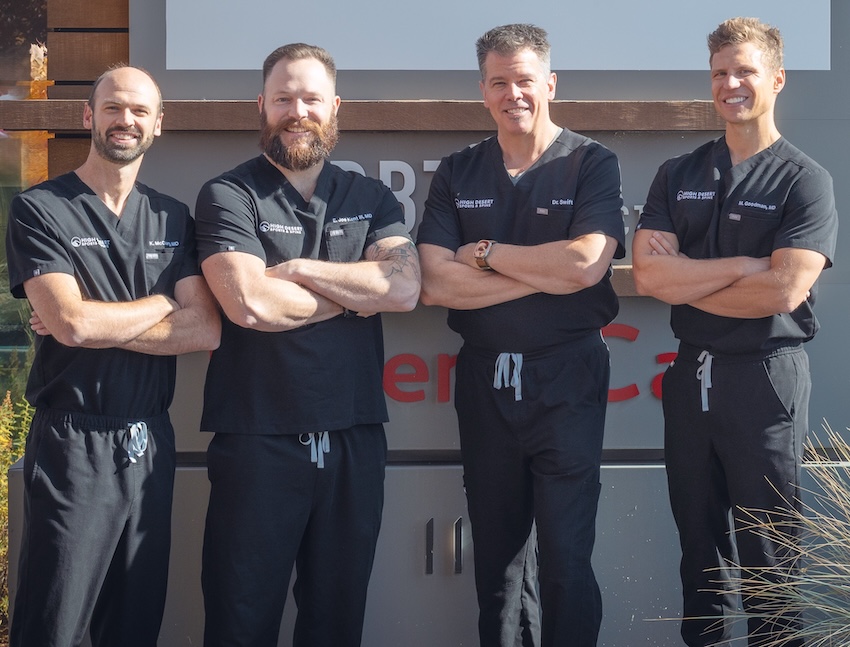
Our physicians completed fellowship training in sports medicine, musculoskeletal medicine, or interventional spine care after medical school and residency, with a focus on tendon, ligament, joint, and nerve problems throughout the body. That whole-body perspective matters for the foot, because how you load it depends on your hip, your knee, and your training.
Shockwave Therapy for Heel Pain
Shockwave is one of the best-evidenced treatments available for plantar fasciitis, and it is not widely offered in Central Oregon. We have both focused and radial systems, which lets us match the treatment to what the ultrasound actually shows rather than to whichever device is in the room.
Diagnostic Ultrasound in the Room
The foot and ankle sit close to the surface, which makes them ideal for ultrasound. We can measure the plantar fascia, look at the Achilles and posterior tibial tendons, assess ligaments while the ankle moves, and see the nerves. A great deal of foot pain is treated on assumption. We would rather look.
We Stay in Our Lane and Say So
We are not a podiatry practice. Podiatrists handle bunions, hammertoes, nail and skin conditions, diabetic wound care, and custom orthotics best, and Central Oregon has good ones. We diagnose and treat tendon, ligament, joint, and nerve problems in the foot and ankle with interventional tools. Knowing the difference saves you time.
Honest About What the Evidence Shows
Cortisone in the plantar fascia carries real risks. We avoid steroid injection around the Achilles entirely. You will get the actual picture rather than a menu.
Same-Week Access
New patient appointments are typically available within a week. For acute injuries, our Orthopedic Urgent Care offers same-day or next-day evaluation. We serve Bend, Redmond, Sisters, Prineville, La Pine, and Sunriver.
Foot & Ankle Pain Treatment in Bend, Oregon — Schedule an Evaluation
If heel pain, Achilles pain, or an unreliable ankle has outlasted rest, stretching, and inserts, the next step is finding out what is actually wrong with the tissue.
We will examine you thoroughly, image the foot in the room, and walk through your options honestly, including which ones the evidence actually supports.
Same-week appointments are typically available for new patients. We serve Bend, Redmond, Sisters, Prineville, La Pine, Sunriver, and patients throughout Central Oregon.
Frequently Asked Questions About Foot & Ankle Pain in Bend, Oregon
Usually one of three reasons. First, chronic plantar fascia pain involves tissue degeneration rather than inflammation, so treatments aimed at inflammation target the wrong issue. Second, the tissue needs graded loading to rebuild, and complete rest can make it weaker. Third, it may not be plantar fasciitis. A heel fat pad problem, a calcaneal stress fracture, and entrapment of a small nerve near the heel can all cause pain in the same area and may not respond to plantar fascia treatment. An ultrasound evaluation can tell you which situation you have.
It has some of the strongest evidence of any treatment for this condition. Systematic reviews and meta-analyses have found meaningful improvements in both pain and function that persist over time, which is not true of every option marketed for heel pain. It is non-invasive, requires no needles and no downtime, and is typically delivered over a short series of sessions. Whether it is right for you depends on what the ultrasound shows and how long you have had symptoms. Most insurance carriers do not cover shockwave therapy, so our team will review the cost with you beforehand.
It can help in the short term, and it can be genuinely useful in some situations, particularly when pain is severe enough that you cannot begin loading the tissue. The concern is what comes with it. Steroid injection into the plantar fascia is associated with rupture of the fascia and with atrophy of the heel fat pad, the natural cushion under your heel, which does not come back. Because of that, guidance has moved away from treating it as a first-line option. We use it selectively and will explain the tradeoff before you decide.
Longer than most people are told. A recent case caught early and managed with load modification, calf and foot strengthening, and footwear changes may settle in a couple of months. Chronic cases that have been building for a year take longer and often need something that restarts the healing process, such as shockwave or PRP. Tissue remodeling takes months, regardless of the treatment used. We would rather set a realistic expectation than a convenient one.
Shin splints usually cause aching that spreads along a stretch of the inner shin and often eases as you warm up. A stress fracture tends to hurt at one specific point, gets progressively worse rather than improving with warm-up, and eventually hurts with walking or at rest. Early X-rays are frequently normal in stress fractures, which is why they get missed. If your shin pain is worsening run over run, stop running and get it evaluated.
Yes, and it is worth addressing rather than living with. Repeated rolling usually means the ligaments healed loose after previous sprains and the nerve feedback system that senses foot position was disrupted. That second part responds well to structured balance and strength training, which is the primary treatment. We also look for what often gets missed in repeatedly sprained ankles, including peroneal tendon injuries, small fractures, and cartilage damage inside the joint. Ankles that keep giving way despite good rehabilitation may need surgical stabilization.
Partly, and it is worth being precise about what that means. Diabetic peripheral neuropathy itself is a medical condition, and blood sugar control, medication management, and diabetic foot and wound care are handled by your primary care provider, a podiatrist, or a neurologist rather than by us. We also want to be clear that neuropathy cannot be reversed by any treatment we offer. We can help identify whether a focal nerve entrapment, such as tarsal tunnel syndrome, is contributing to your symptoms. We offer symptom management with neuromodulation and medical laser, both of which have evidence in managing pain related to diabetic peripheral neuropathy
No. We do not provide or fit custom orthotics. We address footwear and how you are loading the foot as part of the plan, and we refer to podiatry when custom orthotics are the right call. Central Oregon has good podiatrists for that.
Health insurance does not cover PRP, so it is an out-of-pocket expense. Coverage varies by payer and continues to evolve. Contact your insurer directly for current information on your plan. Most insurance carriers do not cover shockwave therapy treatment. Our team can discuss self-pay options during your consultation, and some patients use HSA or FSA funds.
No referral is required. Contact our office directly to schedule an evaluation. Bring any prior imaging or operative reports, and if your pain is related to running or hiking, bring the shoes you have been training in.